Department of Biochemistry
Research theme: Understanding the rules of life
Biography
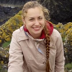
I am from Estonia, where I have lived for the first 19 years of my life and graduated from high school. In 2013, I moved to Edinburgh to study Biochemistry. During this time, I have worked with Dr Meriem El Karoui on DNA damage response in bacteria, and with Dr Sara Buonomo on DNA repair in mouse embryonic stem cells. I have also spent a year as an exchange student in Singapore, which has been an amazing experience. Finally, I've arrived in Cambridge in 2017 to do my PhD, and I am now part of Prof Ernest Laue's group, applying single-molecule microscopy methods to study chromatin structure. Aside from science, I have danced all my life, and currently I am part of the Cambridge University Ballet Club. I also enjoy reading and walking a lot.
Research
Project Title:
Single-molecule microscopy reveals details of heterochromatin reorganisation at the onset of mouse ES cell differentiation
Project Summary:
In the last decade, spatial organisation of the genome has been recognised as one of the important factors linked to gene expression and cell identity. Although we have come a long way in describing how chromatin folds, it is still unclear what determines this structure and why it changes upon differentiation.
Mouse embryonic stem cells provide an excellent system to address these questions, since upon exit from naive pluripotency their genome undergoes a dramatic structural reorganisation. I employ single-molecule microscopy methods to study heterochromatin structure and dynamics in live mESCs and at the onset of their differentiation, focussing on the role of heterochromatin protein 1β (HP1β). By applying novel analysis methods, I test different biophysical models for heterochromatin compartmentalisation in mESCs.
Furthermore, I am developing super-resolution microscopy approaches to image chromatin-binding proteins and DNA in 3D throughout the entire nuclear volume of fixed cells. The protocol is designed to be compatible with single-cell Hi-C, enabling modeling of the genome structures of the imaged cells. Correlating the images and the structures will for the first time make it possible to obtain spatial sequence-specific information about the genome fold alongside protein localisation on a single-cell level. I hope that this method will shed light on the molecular functions of chromatin-binding proteins in their native context and help us understand the rules of genome organisation.
Publications
Basu et al, "Live-cell 3D single-molecule tracking reveals how NuRD modulates enhancer dynamics", 2021, bioRxiv 2020.04.03.003178; doi: https://doi.org/10.1101/2020.04.03.003178
Teaching and Supervisions
Professor Ernest Laue


